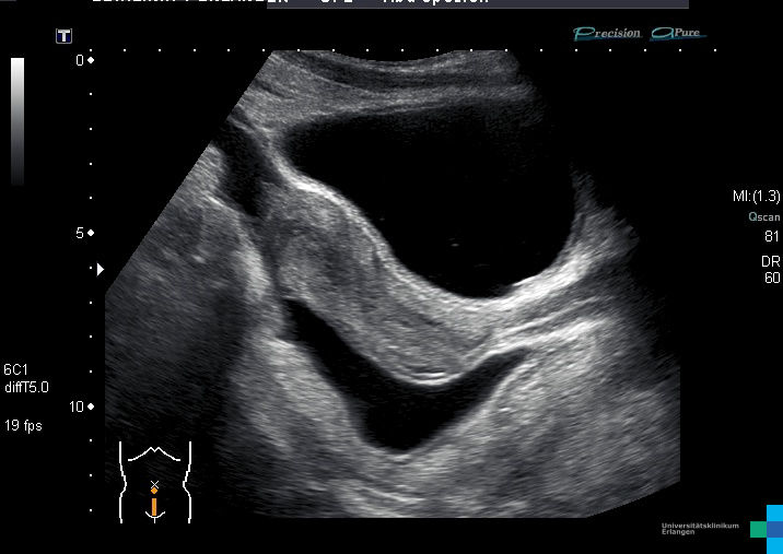
Understanding the Pouch of Douglas on ultrasound is essential for diagnosing and managing various medical conditions. This anatomical structure, located in the female pelvis, plays a crucial role in reproductive health. By leveraging ultrasound technology, healthcare professionals can detect abnormalities and ensure timely interventions. In this article, we will delve into the intricacies of the Pouch of Douglas, its significance in medical imaging, and how it impacts women's health.
The Pouch of Douglas, also known as the rectouterine pouch, is a vital anatomical landmark that is often assessed using ultrasound. As advancements in medical imaging continue, the importance of understanding this structure cannot be overstated. Healthcare providers rely on ultrasound to identify potential issues that could affect fertility, pregnancy, and overall reproductive health.
This article aims to provide a detailed overview of the Pouch of Douglas, its role in ultrasound imaging, and its relevance to clinical practice. By exploring the anatomy, diagnostic techniques, and associated conditions, we aim to equip readers with a comprehensive understanding of this critical topic. Let's dive deeper into the world of ultrasound and the Pouch of Douglas.
Read also:Spruill Oaks Library Your Gateway To Knowledge In Johns Creek Ga
Table of Contents
- Anatomy of the Pouch of Douglas
- Ultrasound Technique for Pouch of Douglas
- Clinical Significance of the Pouch of Douglas
- Common Conditions Detected via Ultrasound
- Diagnosis Using Ultrasound
- Treatment Options for Related Conditions
- Advancements in Ultrasound Technology
- Limitations of Ultrasound in Assessing the Pouch of Douglas
- Recent Research and Studies
- Conclusion and Call to Action
Anatomy of the Pouch of Douglas
The Pouch of Douglas is an anatomical structure located in the female pelvis, specifically between the rectum and the posterior wall of the uterus. It is part of the peritoneal cavity and serves as a potential space that can accumulate fluid in certain pathological conditions. Understanding its location and function is essential for healthcare providers when interpreting ultrasound images.
Key Features of the Pouch of Douglas
- Located posterior to the uterus
- Forms the lowest point of the female peritoneal cavity
- Can accumulate fluid, blood, or pus in pathological conditions
This anatomical structure is named after Dr. James Douglas, a Scottish anatomist who first described it. Its position makes it a critical point of focus during pelvic ultrasound examinations, particularly in cases involving fluid accumulation or mass detection.
Ultrasound Technique for Pouch of Douglas
Ultrasound imaging is a non-invasive, safe, and effective method for evaluating the Pouch of Douglas. The technique involves using high-frequency sound waves to create detailed images of the pelvic structures. This section will explore the specific steps and considerations involved in conducting an ultrasound examination of the Pouch of Douglas.
Steps in Performing an Ultrasound
- Position the patient comfortably on the examination table
- Use a transabdominal or transvaginal ultrasound probe
- Ensure adequate visualization of the pelvic structures
Transvaginal ultrasound is often preferred for better resolution and clarity, especially when assessing the Pouch of Douglas. The use of color Doppler imaging can further enhance the evaluation of blood flow and fluid dynamics in this region.
Read also:Discovering Walgreens Waite Park Mn A Complete Guide To Services Products And More
Clinical Significance of the Pouch of Douglas
The Pouch of Douglas holds significant clinical importance due to its role in various gynecological conditions. Its anatomical position makes it a common site for fluid accumulation, which can indicate underlying pathologies such as endometriosis, pelvic inflammatory disease, or ovarian cysts. Recognizing these conditions early through ultrasound can lead to timely interventions and improved patient outcomes.
Conditions Related to the Pouch of Douglas
- Endometriosis
- Pelvic inflammatory disease (PID)
- Ovarian cysts
- Ectopic pregnancy
Healthcare providers must be vigilant in identifying any abnormalities in the Pouch of Douglas during routine ultrasound examinations. Early detection and management of these conditions are crucial for maintaining reproductive health.
Common Conditions Detected via Ultrasound
Ultrasound plays a pivotal role in diagnosing various conditions associated with the Pouch of Douglas. By providing detailed images of the pelvic structures, ultrasound can help identify fluid accumulation, masses, and other abnormalities. This section will discuss some of the most common conditions detected using this technology.
Endometriosis
Endometriosis is a condition where tissue similar to the lining of the uterus grows outside the uterine cavity. The Pouch of Douglas is a frequent site for endometrial implants, leading to pain and infertility. Ultrasound can detect these implants and guide treatment decisions.
Pelvic Inflammatory Disease
Pelvic inflammatory disease (PID) is an infection of the female reproductive organs that can cause fluid accumulation in the Pouch of Douglas. Ultrasound is instrumental in diagnosing PID and monitoring treatment progress.
Diagnosis Using Ultrasound
Diagnosing conditions related to the Pouch of Douglas requires a thorough understanding of ultrasound techniques and interpretation. Healthcare providers must be skilled in recognizing normal and abnormal findings during these examinations. This section will outline the key steps involved in diagnosing conditions using ultrasound.
Key Steps in Diagnosis
- Assess the size and shape of the Pouch of Douglas
- Evaluate for fluid accumulation or masses
- Use Doppler imaging to assess blood flow
Combining ultrasound findings with clinical symptoms and other diagnostic tests can enhance the accuracy of diagnosis. Collaboration between healthcare providers and radiologists is essential for optimal patient care.
Treatment Options for Related Conditions
Once a condition related to the Pouch of Douglas is diagnosed, appropriate treatment options can be explored. The choice of treatment depends on the specific condition and its severity. This section will discuss some of the most common treatment approaches for conditions associated with the Pouch of Douglas.
Surgical Interventions
In cases where fluid accumulation or masses are detected, surgical intervention may be necessary. Laparoscopy is a minimally invasive procedure that allows for the removal of endometrial implants or other abnormalities. This approach offers quicker recovery times and fewer complications compared to traditional surgery.
Medications
Medications such as antibiotics, hormonal therapies, and pain relievers are often used to manage symptoms and treat underlying conditions. The choice of medication depends on the specific diagnosis and patient preferences.
Advancements in Ultrasound Technology
Recent advancements in ultrasound technology have significantly improved the ability to assess the Pouch of Douglas and surrounding structures. High-resolution imaging, three-dimensional (3D) ultrasound, and elastography are just a few examples of innovations that have enhanced diagnostic capabilities. This section will explore these advancements and their impact on clinical practice.
Three-Dimensional Ultrasound
Three-dimensional ultrasound provides detailed images of the pelvic structures, allowing for better visualization of the Pouch of Douglas and its surroundings. This technology can help detect subtle abnormalities that may be missed with traditional two-dimensional imaging.
Limitations of Ultrasound in Assessing the Pouch of Douglas
While ultrasound is a powerful tool for evaluating the Pouch of Douglas, it does have certain limitations. Factors such as patient obesity, bowel gas, and operator skill can affect image quality and diagnostic accuracy. This section will discuss these limitations and their implications for clinical practice.
Impact of Patient Factors
Patient factors such as obesity and bowel gas can obscure the view of the Pouch of Douglas during ultrasound examinations. In such cases, alternative imaging modalities like MRI may be necessary for a more comprehensive assessment.
Recent Research and Studies
Ongoing research continues to expand our understanding of the Pouch of Douglas and its role in various medical conditions. Studies exploring new imaging techniques and treatment options are helping to improve patient outcomes. This section will highlight some of the latest research findings in this field.
Emerging Imaging Techniques
Emerging imaging techniques such as shear wave elastography and contrast-enhanced ultrasound are showing promise in improving the detection and characterization of abnormalities in the Pouch of Douglas. These technologies offer enhanced sensitivity and specificity, leading to more accurate diagnoses.
Conclusion and Call to Action
In conclusion, the Pouch of Douglas plays a critical role in women's health, and its assessment through ultrasound is essential for diagnosing and managing various conditions. By understanding its anatomy, clinical significance, and associated conditions, healthcare providers can ensure timely interventions and improved patient outcomes.
We encourage readers to share this article with others who may benefit from the information provided. Additionally, we invite you to explore other articles on our site that delve into related topics in women's health. Together, we can promote awareness and advance the field of medical imaging.
For further reading and references, please consult the following sources:
- ACOG Practice Bulletin on Endometriosis
- Royal College of Obstetricians and Gynaecologists Guidelines
- Recent studies published in the Journal of Ultrasound in Medicine


